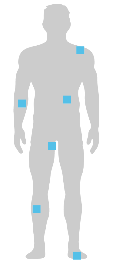
Three-dimensional solution for deep and tunneling wounds

New HCPCS Level II Code A2025
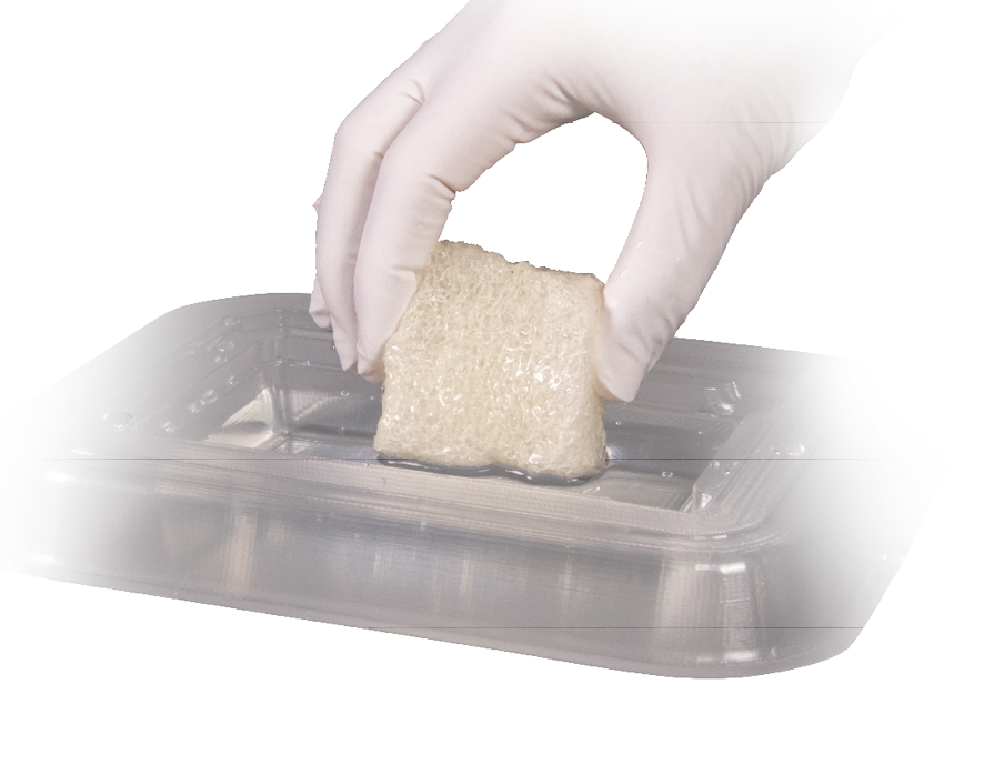
Miro3D is designed for use in deep and tunneling wounds

  • 2cm thick collagen sheet scaffold designed for deep, tunneling and irregular wounds
  • Open and porous acellular graft provides protective environment for wound management
  • Cut to desired size for full wall apposition and conformity to wound beds

Unlike thin grafts, Miro3D provides volume in deep wounds

Broad range of clinical uses

  • Stage III and IV pressure ulcers
  • Necrotizing fasciitis
  • Partial and full-thickness wounds
  • Wound dehiscence
  • Perirectal abscess
  • Pilonidal sinus disease
  • Diabetic foot ulcers
  • Trauma wounds
  • Other tunneled and undermined wounds

Clinical Case Studies*

Foot Abscess Treated With Miro3D Wound Matrix

Clinical Overview and History

Male patient presented in the emergency department with cellulitis of the left foot. The patient noticed an increase in redness, warmth, swelling, and pain (despite neuropathy) of the left foot, two weeks prior to visiting the hospital.

Health history includes diabetes mellitus, neuropathy, high blood pressure, and a deformity of the left foot due to a previous gunshot wound.

Initial Findings

A CT scan of the left foot showed inflammatory changes consistent with cellulitis but without evidence osteomyelitis. Other signs include edema, redness, warmth, and swelling. A wound of the foot was noted to have a foul odor with drainage (Figure 1).

Treatment

The patient was brought into the operating room for incision and drainage of the left foot abscess. An approximate 17cm wound remained following the removal of pus and necrotic tissue (Figure 2). The wound was flushed with Irrisept® and packed with Aquacel® Ag Advantage and a dry dressing. A wound VAC was applied the next day and IV antibiotics were started.

Six days later, the patient returned to the operating room and an ongoing infection was noted, including another abscess near the left ankle. The abscess was drained, increasing the incision by 6cm (Figure 3). The wound was flushed again with Irrisept®, packed with Hema-Flex Sealant (due to bleeding) and Aquacel® Ag Advantage and a dry dressing. The wound VAC was reapplied.

Post 11 days from the first visit, the patient was discharged with the wound VAC and IV antibiotics. The wound measured 21.5cm x 4cm x 2cm.

One week later, the patient was seen in the office where the wound VAC was removed (Figure 4). Aquacel® Ag Advantage was applied again and the patient was instructed to perform a dressing change and continue IV antibiotics prior to surgery.

Four days later, over 3 weeks from the first visit, the patient was brought into the operating room for surgical debridement and application of Miro3D wound matrix. The wound measurements were 22cm x 4cm x 1.5cm (Figure 5). Three Miro3D (Two 10cm x 5cm x 2cm and one 2cm x 2cm x 2cm) units were packed into the deep wound (Figures 6). A Mepitel® dressing was placed directly over the Miro3D. A wound VAC at 125mmHg continuous with black foam was applied.

Post three days from the placement of Miro3D, the wound measured 21cm x 3.8cm x 0.4cm (Figure 7), demonstrating a significant improvement in depth of the wound. No debridement was performed, and the wound VAC was reapplied at 125mmHg continuous. The patient continued IV antibiotics.

At follow up, seven days from initial application of Miro3D, the measurements of the wound were 21cm x 3.4cm x 0.3cm (Figure 8). No debridement was performed, and both wound VAC therapy and IV antibiotics were continued.

Post day 10 from Miro3D placement, the wound measured 21cm x 3.1cm x 0.1cm with one area measuring 0.2cm in depth (Figure 9). The patient continued IV antibiotics and wound VAC therapy.

The wound measured 20.2cm x 2.6cm x 0.1cm, post 17 days from placement of Miro3D (Figure 10). The patient continued IV antibiotics and wound vac therapy. Four days later, the wound measured 19cm x 2.8cm x 0.1cm. Miro3D Fibers (500mg) were applied to the wound. IV antibiotics and wound vac therapy were continued.

On day 25 from the initial placement of Miro3D and 4 days from the application of Miro3D Fibers, the measurements of the wound were 18.6cm x 2.8cm x 0.1cm (Figure 11). The patient continued IV antibiotics and wound vac therapy.

The patient continued with IV antibiotics and wound vac therapy while following up in the clinic. The wound continued to decrease in size with the following measurements and time-points from the initial placement of Miro3D Fibers:

Post application of Miro3D Post application of Miro3D Fibers Wound Measurements Figure
35 days 14 days 17.5cm x 2.8cm x 0.3cm 12
42 days 21 days 9.2cm x 2.8cm x 0.2cm 13
49 days 28 days 8cm x 1cm x 0.1cm 14
56 days 35 days 5cm x 1cm x 0.1cm 15
61 days 42 days 1cm x 0.6cm x 0.1cm 16
68 days 49 days 0.8cm x 0.4cm x 0.1cm 17

Summary

Miro3D wound matrix is designed to provide wound bed conformity in deep, irregular wounds, which was impressively demonstrated in this case. Within three days of placing Miro3D, the physician observed significant integration of Miro3D with the depth of the wound decreased by over 70%. The overall wound size continued to steadily decrease over 68 days after only one application of Miro3D and one application of Miro3D Fibers. The patient will receive weekly standard dry dressing changes with a complete closure of the wound expected within the next seven to ten days.

Physician Information

Raymond John Abdo, DPM, Mercy Hospital South, St. Louis, Missouri
Dr. Raymond Abdo DPM is a foot and ankle surgeon in Saint Louis, MO specializing in Foot & Ankle Surgery and Podiatry (Foot & Ankle Medicine). Dr. Abdo is also in private practice at locations in St. Louis and Eureka, MO. He graduated from Des Moines University College of Podiatric Medicine and Surgery in 2002 and has 19 years of experience. Raymond Abdo DPM is affiliated with Mercy Hospital – St. Louis and Mercy (Arkansas, Kansas, Missouri, and Oklahoma).

Amy Louise Couch, MD, Mercy Hospital South, St. Louis, Missouri
Dr. Amy Couch is a board certified family medicine physician specializing in wound care and hyperbaric medicine. She received her medical degree from the six-year combined BA/MD program at the University of Missouri-Kansas City graduating Summa Cum Laude in 2007 and inducted into Alpha Omega Alpha Medical Honor Society and Gold Humanism Honor Society. She did an internship in Obstetrics and Gynecologic Surgery and she completed her Family Medicine residency at the Mayo Clinic in Rochester, MN, where she served as chief resident. She practiced as an urgent care physician for 10 years at Mercy Clinic where she also had a medical director role. She currently serves as the medical director at Mercy South Hyperbaric & Wound Center in St. Louis, MO.

Foot Abscess Treated With Miro3D Wound Matrix

Clinical Overview and History

50 year old female with a history of diabetes who presented in the emergency department with cellulitis and ulceration sub right first metatarsophalangeal joint (MPJ). The patient had been treating the ulceration with Betadine® and using a controlled ankle motion (CAM) walker boot for 2 weeks prior to the visit.

Health history includes diabetes mellitus, neuropathy, high blood pressure, high cholesterol, hypothyroidism, and a deformity of the right foot.

Initial Findings

Labs indicated WBC = 9.5, anemia, elevated glucose, Cr = 0.89. Radiographic imaging showed no evidence of osteomyelitis. MRI results showed subcutaneous fluid thought to be an abscess at plantar 1st MPJ and mild edema noted within lateral sesamoid.

The ulceration contained odorous, serosanguinous drainage and undermining with necrotic tissue. Cellulitis was present along the periphery of the ulceration.

Treatment

Four days later, the patient was brought into the operating room for incision and drainage of the right foot abscess with right lateral sesamoidectomy. Silver Alginate was applied along with a dry dressing. A wound VAC applied and utilized during the five day hospital stay.

The patient was prescribed IV antibiotics upon discharge from the hospital but the wound VAC was discontinued due to insurance coverage challenges.

Approximately two and half weeks from the original incision and drainage surgery, the patient returned for outpatient surgical debridement using a VERSAJET Hydrosurgery System. The wound measured 2cm x 2.2cm x 2cm and contained moderate serosanguineous drainage (Figure 1). Following debridement, a Miro3D (3cm x 3cm x 2cm) wound matrix was applied followed by a nonadherent dressing, Steri-Strips, Silver Aginate and dry dressings (Figure 2).

Post 35 days from the Miro3D placement, the diameter of the wound measured 1.5cm x 1.5cm with 0.5cm in depth (Figure 3). No odorous drainage was present. Fibrous tissue and slough were noted. The wound was debrided using a currette and Miro3D (3cm x 3cm x 2cm) was placed for the second time (Figure 4). Adaptic™, Steri-Strips™, Silver Alginate, and dry dressings were used to secure Miro3D in place.

At follow up, three days following the second placement of Miro3D, the measurements of the wound were 1.4cm x 1.1cm x 0.3cm (Figure 5). No debridement was performed. Adaptic™, Silver Alginate, and dry dressings were utilized.

One week later, the wound measurements on the right foot were 1.2cm x 1.1cm x 0.3cm. A Total Contact Cast (TCC) was applied with collagen, Hydrofera Blue(R) dressing, and an ABD with Kerlix gauze dry dressing. The patient was prescribed oral antibiotics long term for suppression.

The patient continued to attend weekly follow up visits at the wound care center. Each visit the wound was debrided, and standard dressings were applied with the TCC. The following are the wound measurements at each follow up time-points from the second application of Miro3D:

  • Post 31 days the wound measured: 1cm x 0.8cm x 0.1cm (Figure 6).
  • Post 38 days the wound measured: 0.8cm x 0.6cm x 0.1cm (Figure 7).

Conclusion and Post Procedure

After one final week in the TCC, the wound was fully healed post 52 days following the second application of Miro3D, 87 days from the initial application of Miro3D (Figure 8).

Summary

The 2cm thickness of Miro3D allowed for full wall apposition and conformity to this deep wound bed. Along with proper off-loading, Miro3D provided a protective environment enabling the wound to achieve full healing within 12 weeks after only two applications of Miro3D.

Physician Information

Raymond John Abdo, DPM, Mercy Hospital South, St. Louis, Missouri
Dr. Raymond Abdo DPM is a foot and ankle surgeon in Saint Louis, MO specializing in Foot & Ankle Surgery and Podiatry (Foot & Ankle Medicine). Dr. Abdo is also in private practice at locations in St. Louis and Eureka, MO. He graduated from Des Moines University College of Podiatric Medicine and Surgery in 2002 and has 19 years of experience. Raymond Abdo DPM is affiliated with Mercy Hospital – St. Louis and Mercy (Arkansas, Kansas, Missouri, and Oklahoma).

Abdominal Surgical Dehiscence Post Panniculectomy With Miro3D Applied to Wound

Clinical Overview and History

Female patient who underwent a panniculectomy surgery in May 2022.

Initial Findings

Approximately two and half months later, the patient presented with an abdominal surgical wound dehiscence. The wound measured 8cm x 2cm x 0.5cm in depth with 2cm of medial tunneling.

A wound VAC was applied. The wound VAC dressing was disrupted and not repositioned which resulted in superficial skin breakdown on the superior part of the wound opening. The VAC was removed by the patient before the two week follow-up visit.

Treatment

Two weeks later, and because of the discontinued use of the wound VAC by the patient, the main wound measured 9cm x 2.7cm x 2cm in depth with 2.5cm of medial tunneling (Figure 1). Miro3D (5cm x 5cm x 2cm size) was applied to the tunneling space and the main body wound (Figure 2). The Miro3D was covered with Mepitel® dressing and secured with an ABD dressing. The patient was instructed to perform daily outer dressing changes and Vashe® wound solution rinses to keep the wound clean. A wound VAC was not reapplied.

Seven days following the initial placement of Miro3D, the wound measured 7.5cm x 2.5cm x 0.5cm in depth with no tunneling (Figure 3). The Miro3D was resorbed into the wound bed. Daily wet to dry dressing changes were initiated.

Three days later, the patient returned for a second application (Figure 4). A 5cm x 5cm x 2cm Miro3D was reapplied to the wound.

Four days after the second placement of Miro3D, the wound measured 7.25cm x 1.25cm x 0.5cm deep and Miro3D was noted as fully resorbed within the wound bed (Figure 5). The wound was washed with a wound solution and covered with a dry dressing. The patient was instructed to perform daily Vashe washes with dry dressing changes. The patient returned for follow-up appointments every one to three weeks as transportation resources allowed.

By day 54, following the second placement of Miro3D, the wound measured 2cm x 1cm x 0.4cm (Figure 6).


77 days from the second placement of Miro3D, observations of the wound included a significant reduction in size and continued wound healing (Figure 7).
On day 98, the wound measured 0.4cm x 0.4cm x 0.2cm (Figure 8).

Conclusion and Post Procedure

Full wound healing was observed in January 2023, which was 127 days from the initial Miro3D application to the wound (Figure 9).

Summary

In this case, the ability to cut and shape Miro3D to conform to the wound bed for full wound wall apposition in both the tunneling space and main wound bed was beneficial. Miro3D demonstrated volume replacement by quickly eliminating the initial tunneling area as well as reducing the size of the wound with the protection of the collagen scaffold. Full healing was achieved 127 days following the placement of Miro3D.

Physician Information

Moses K. Shieh, DO, FACOS, Medical Director & Founder of Surgical Healing Arts Center in Fort Myers, FL
Specializes in Advanced Laparoscopic Bariatric Surgery and General Surgery. Surgical Director of Robotic Surgery at Gulf Coast Medical Center (part of the Lee Health System) in Fort Myers and currently serves as a member of the Medical Executive committee. Wound Director for Landmark Hospital in Naples.

Necrotizing Fasciitis Resulting in Multi-Compartment Wound with Tunneling Treated with Miro3D Wound Matrix and MiroDerm Biologic Wound Matrix

Clinical Overview and History

Female patient with a history of bipolar disorder and arthritis, presented to the emergency room with complaints of pain in the left groin area for the past 24 hours.

Initial Findings

Upon physical examination, an area of induration, erythema, and tenderness were noted in the left groin and lateral to the labia.

A CT scan suggested a necrotizing soft tissue infection with gas in the perilabial region and left lower quadrant. Additionally, the patient’s white blood cell count was 28.4.

Treatment

The patient was transferred to the operating room for debridement. The next day, additional debridement was performed as well as two days later, resulting in multiple wounds (Figure 1).

Wound #1
Superiorly to the groin, the first wound measured 10cm x 5cm x 5cm with an additional 5cm x 4cm x 2cm undermining pocket located laterally in the subcutaneous space (Figure 2). A subcutaneous tunnel measuring 6cm x 2cm x 2cm connected the superior groin wound to the second wound located lateral to the labia.

The following Miro3D units were used to fill this significant wound space:

  • Superior groin wound: Miro3D Wound Matrix 10cm x 5cm x 2cm
  • Subcutaneous pocket wound: Miro3D Wound Matrix 5cm x 5cm x 2cm
  • Tunneling wound: Miro3D Wound Matrix 3cm x 3cm x 2cm

Negative Pressure Wound Therapy (NPWT) was applied at 50mmHg with intermittent medium suction.

Initial resorption of Miro3D was observed within seven days.

Post 14 days from the application of Miro3D, the wounds were irrigated and the following measurements were documented:

  • Superior groin wound: 10cm x 2cm x 1cm – 92% reduction in total volume with an 80% decrease in overall depth of the wound.
  • Subcutaneous pocket wound: 5cm x 2cm x 1cm – 75% reduction in total volume of the wound with a 50% decrease in depth.
  • Tunneling wound: 5cm x 2cm x 1cm – 41% decrease in total volume.

At the initial 14-day follow-up visit, Miro3D (10cm x 5cm x 2cm and 3cm x 3cm x 2cm) units were reapplied to the superior groin and subcutaneous pocket wound. The patient was discharged the following day with NPWT and a follow up appointment scheduled in the wound care center.

At follow up, post 15 days following the second application, the subcutaneous pocket and tunneling wound were closed. The superior groin wound measured 6.8cm x 1cm x 0.5cm a 99% reduction in the total volume from the initial wound size.

Post 36 days from the second application, the superior groin wound continued to decrease, measuring 5cm x 0.8cm x 0.5cm (Figure 3).

Post 50 days from the second application of product, the superior groin wound treated with Miro3D was completely healed.

Wound #2
The second wound, located lateral to the labia, initially measured 15cm x 8cm x 4cm. MiroDerm Fenestrated (8cm x 15cm size) was applied (Figure 4) with NPWT.

Post 14 days from the application of product, the perilabial wound measured 8cm x 4cm x 3cm. One additional application of MiroDerm Fenestrated Plus (8cm x 8cm) and NPWT were reapplied.

The perilabial wound continued to decrease with the following measurements and time points from the second application:

  • Post 15 days wound measured: 6cm x 3cm x 0.4cm
  • Post 36 days wound measured: 6cm x 0.5cm x 0.4cm (Figure 5)

Post 50 days from the second MiroDerm application, the perilabial wound measured 0.8cm x 0.3cm x 0.1cm.

Post 64 days from the second application of product, the perilabial wound treated with MiroDerm was fully healed.

Conclusion and Summary

This complicated and massive multi-compartment necrotizing fasciitis wound fully healed in 11 weeks after only two applications of Miro3D and MiroDerm in each wound respectively. Miro3D wound matrix impressively demonstrated the benefits of its porous nature and conformability to deep wound beds, and large undermining and tunneling wound spaces by significantly reducing the depth of the superior groin wound by 80% within 14 days, after the first application of product.

The subcutaneous pocket wound was reduced by 50% within the same time frame and after the first application of Miro3D. Additionally, following the second application, the wound was fully healed within 10 days.

The three-dimensional structure of the Miro3D appears to be a valuable tool when faced with treating large undermining and tunneling spaces. It would be challenging to get a flat graft to conform to these dead spaces.

MiroDerm biologic wound matrix also successfully managed the large area of the perilabial wound by decreasing the area by 74% within 14 days of the initial placement of product, with full wound healing noted at 64 days.

Physician Information

Brian Parkes, MD, FACS, Medical Director of Scotland Wound Healing, Scotland Memorial Hospital, Laurinburg, North Carolina

Pilonidal Sinus Abscess Successfully treated with Miro3D

Clinical Overview and History

Patient is a male with a history of recurrent pilonidal cyst infection (spontaneous drainage) with use of oral antibiotics.

Initial Findings

  • Presented in September 2022 with pilonidal cyst recurrence
  • Surgical consult conducted to perform excision of cyst

Treatment

On September 21, 2022, patient underwent a pilonidal cyst surgical excision without closure. Post excision, the wound measured 4cm x 3cm x 2.5cm in depth. A wound VAC with white foam dressing was applied at 125mmHg for 5 days post excision (Figure 1).

Post day 5, Miro3D (5cm x 5cm x 2cm size) was placed and secured using a full thickness suture technique (Figure 2). A wound VAC, using white foam dressing, was applied at 125mmHg for an additional 6 days.

Six days later, the wound VAC was removed. Wound site observation included complete incorporation of Miro3D and ingrowth of granulation tissue (Figure 3).

Over the next three weeks, daily moist to dry dressing changes were performed.

Figure 4 demonstrates the healing progression observed on week 2 of the 3 total weeks of daily dressing changes.

Conclusion and Post Procedure

After three weeks of dressing changes, post 29 days from the initial Miro3D application to the wound, observation of 100% wound healing without deformity was noted (Figure 5).

Summary

Miro3D quickly integrated into the native bed and within 7 days from placement of Miro3D, complete incorporation and ingrowth of granulation tissues was observed. Full healing was observed without deformity 29 days after placement of Miro3D.

From past experience in surgical excisions of pilonidal sinus abscess cases, I would anticipate this extent of healing only after approximately 3-4 months of treatment using daily dressing changes and weekly debridement.

Physician Information

Rodney Miller, MD, General Surgery, Crossroads Providers
Board-certified general surgeon with specialized training in robotic surgery. Dr. Miller obtained his medical degree and completed his general surgery residency at East Tennessee State University and has over 25 years of experience. Dr. Miller completed his fellowship in Laparoscopic Colectomy Hands-On from Washington University, a fellowship in Healogics Advanced Wound Care and Hyperbaric Oxygen Therapy in Destin Florida, and an Advanced Laparoscopic Nissen Education Curriculum from the University of Nebraska Medical Center School of Medicine.

Large Sebaceous Cyst Excised And Successfully Treated With Miro3D Wound Matrix

Clinical Overview and Initial Findings

Patient presented to the clinic for a scheduled pre-operative appointment prior to a planned hernia repair surgery.

Upon the perioperative visit, the patient was found to have an infected, draining sebaceous cyst in the upper scapular region. Due to the discovery, the hernia repair was postponed. The patient was started on oral antibiotic therapy for 14 days prior to excision of the cyst.

Treatment

The sebaceous cyst was excised leaving a wound size of 11cm x 7cm (Figure 1). Miro3D wound matrix (10cm x 5cm x 2cm) was placed onto the wound with WHITEFOAM™ and a wound VAC 125mmHg.

Five days post excision and after placement of Miro3D and a wound VAC, small visual residual amounts of Miro3D was observed within the wound bed (Figure 2). The wound bed was left undisturbed and not debrided at this time to allow any residual Miro3D to continue to incorporate. The wound was dressed with a non adherent and secured in place with a standard secondary dressing.

Ten days later, post 15 days from the initial Miro3D application, the wound measured 9cm x 5cm (Figure 3). Standard care of debridement and dressings were performed.

Post 22 days from excision and placement of Miro3D, healthy granulation tissue was observed within the wound bed (Figure 4).

Due to prolific granulation tissue (Figure 5), silver nitrate was applied post 36 days from excision and the placement of Miro3D (Figure 6). The physician was pleased with the continued healing process and indicated he would rather apply an antidote to prolific granulation overgrowth vs. be challenged with minimal granulation tissue growth and wound stalling.

By 43 days, the wound measured 7cm x 3cm (Figure 7). Silver nitrate was applied a second time due to hypergranulation tissue.

The patient continued with dry dressing changes as needed and followed up in the clinic weekly. The wound continued to decrease in size with the following measurements and timepoints from excision and placement of Miro3D:

  • Post 56 days wound measured: 5cm x 2.5cm (Figure 8)
  • Post 63 days wound measured: 3.5cm x 1.5cm (Figure 9)
  • Post 83 days wound measured: 1.2cm x 0.8cm (Figure 10)

Conclusion and Post Procedure

Full wound healing was achieved on day 97 (Figure 11).

Summary

Due to the highly porous structure of Miro3D, it is able to conform well to the intended space and quickly integrate into the wound bed providing a protective environment for wound management. This quick integration was demonstrated post 5 days from placement, allowing the wound to fully heal within 97 days after only one application of Miro3D. Subsequently, the patient was able to undergo a successful hernia repair surgery on day 97.

Physician Information

Rodney Miller, MD, General Surgery, Crossroads Providers 
Board-certified general surgeon with specialized training in robotic surgery. Dr. Miller obtained his medical degree and completed his general surgery residency at East Tennessee State University and has over 25 years of experience. Dr. Miller completed his fellowship in Laparoscopic Colectomy Hands-On from Washington University, a fellowship in Healogics Advanced Wound Care and Hyperbaric Oxygen Therapy in Destin Florida, and an Advanced Laparoscopic Nissen Education Curriculum from the University of Nebraska Medical Center School of Medicine.

*The experience depicted in these case studies may not be representative of all potential clinical outcomes. Results may vary by patient. Further study is necessary to determine the benefit of Miro3D in a variety of wound applications.

Available in four sizes that can be cut and shaped to fill and maintain direct contact with irregular wound beds
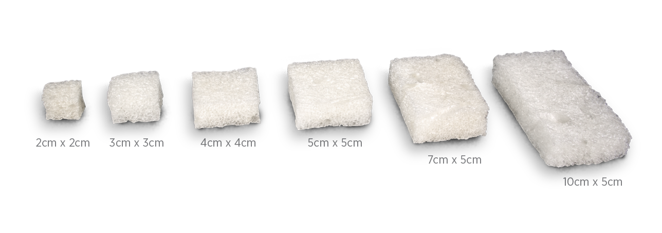
All sizes = 2cm thickness

Ordering Information

ITEM ID SIZE (cm) TOTAL (cm3)
3000 2 x 2 x 2 8
3005 3 x 3 x 2 18
3010 5 x 5 x 2 50
3015 10 x 5 x 2 100

Packaged 1 per box.

Miro3D wound matrix is indicated for the management of wounds, including: partial and full-thickness wounds; pressure ulcers; venous ulcers; chronic vascular ulcers; diabetic ulcers; tunneled, undermined wounds; trauma wounds (abrasion, lacerations, partial thickness burns, skin tears); drainage wounds; and surgical wounds (donor sites/grafts, post-Mohs surgery, post-laser surgery, podiatric, wound dehiscence).

CAUTION: Federal (USA) law restricts this device to sale by or on the order of a physician.
Refer to the Instructions for Use for a complete listing of the indications, contraindications, warnings and precautions. Information in this material is not a substitute for the product Instructions for Use.

Download IFU here.

Available in a fiber version designed for irregular wound beds
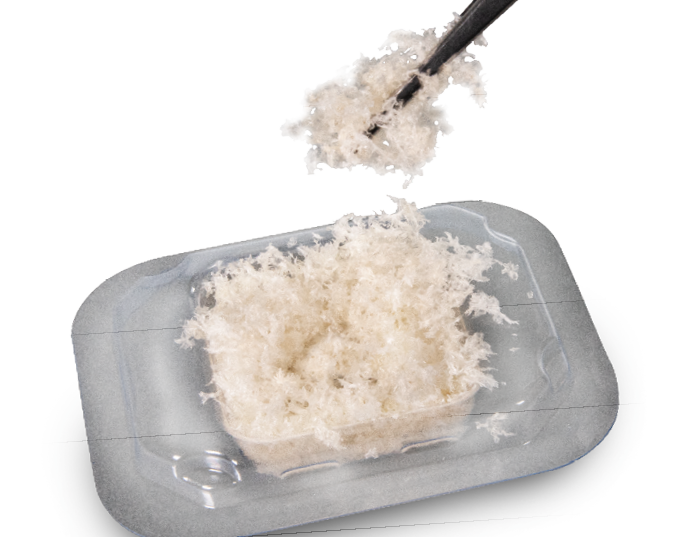
Ordering Information

ITEM ID SIZE (mg)
4000 100
4005 250
4010 500
4015 700

Packaged 1 per box.

Miro3D Fibers wound matrix is intended for the management of wounds including: partial and full thickness wounds, pressure ulcers, venous ulcers, chronic vascular ulcers, diabetic ulcers, tunneled, undermined wounds, trauma wounds (abrasions, lacerations, partial thickness burns, and skin tears), draining wounds, surgical wounds (donor sites/grafts, post-Mohs surgery, post-laser surgery, podiatric, wound dehiscence).

CAUTION: Federal (USA) law restricts this device to sale by or on the order of a physician.
Refer to the Instructions for Use for a complete listing of the indications, contraindications, warnings and precautions. Information in this material is not a substitute for the product Instructions for Use.

Download IFU here.

Product Inquiries and Orders

Call us at: 763-284-6780

Fax us at: 952-856-5085

Product Inquiries at: customerservice@reprisebio.com

SM-00198 Rev. J 08/23